
Non-Invasive Vascular Treatments: Comprehensive Guide to Minimally Invasive Vein Care
- Dec 22, 2025
- 11 min read
Updated: Feb 18
Non-invasive vascular treatments are minimally invasive procedures that repair, close, or remove problematic veins without open surgery, reducing recovery time and procedural risk while restoring function and appearance. This guide explains the common conditions treated non-invasively in Toronto, the principal procedures available locally, and how each approach works, so patients can evaluate options with clinical clarity. Many people with varicose veins, spider veins, venous insufficiency, or vascular lesions want effective care without long hospital stays or general anesthesia; minimally invasive vascular procedures address those needs through targeted energy, chemical agents, or adhesives.
The article walks through diagnosis and imaging, compares Endovenous Laser Treatment (EVLT), Sclerotherapy, Radiofrequency Ablation (RFA) and the VenaSeal Closure System, outlines recovery timelines and lifestyle measures, and explains OHIP coverage realities and practical payment navigation. Throughout, the emphasis is on evidence-informed mechanisms, expected outcomes, and the questions to ask during a consultation in Toronto clinics offering duplex ultrasound–guided care. The goal is to give readers practical next steps for selecting treatment and preparing for recovery in a local, minimally invasive care pathway.

What Are the Common Vascular Conditions Treated Non-Invasively in Toronto?
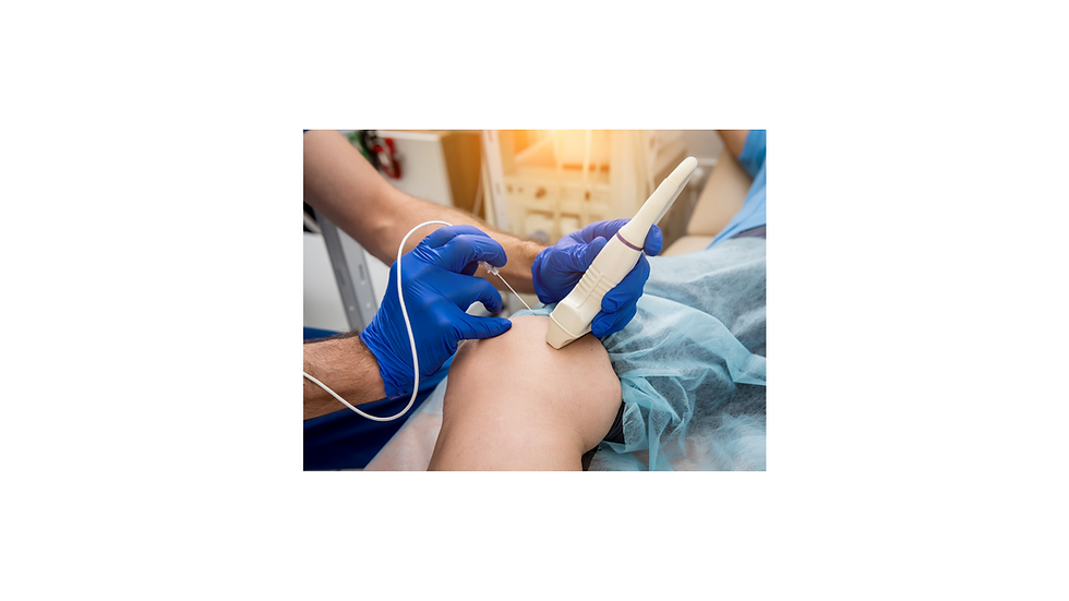
Non-invasive vascular treatments in Toronto target superficial and some deeper venous problems that impair circulation or cause symptoms and cosmetic concerns. These conditions include chronic venous insufficiency leading to varicose veins, spider veins (telangiectasia), localized vascular lesions, recurrent superficial thrombophlebitis, and select situations related to post-thrombotic syndrome where endovascular approaches help restore flow. Each condition has distinct signs—bulging cords and aching for varicose veins, fine red or blue lines for spider veins, and persistent swelling or skin changes for more advanced venous insufficiency—so accurate diagnosis directs the appropriate minimally invasive option. Diagnosing these disorders typically uses a structured history, focused physical exam, and duplex ultrasound to map reflux and anatomy, which informs whether thermal ablation, sclerotherapy, adhesive closure, or a combination is the best approach. Understanding these conditions and their diagnostic pathway helps patients decide when to pursue intervention versus conservative management and prepares them for discussion of targeted procedures.
The most commonly treated vascular conditions include:
Varicose veins that cause pain, heaviness, or skin changes often benefit from vein closure or removal.
Spider veins and small vascular lesions are primarily cosmetic but sometimes symptomatic.
Chronic venous insufficiency presenting with leg swelling, skin discoloration, or ulceration risk requires intervention.
Recurrent superficial thrombophlebitis or symptomatic venous segments where endovascular treatment reduces symptoms and progression.
These categories determine whether treatment focuses on symptom relief, cosmetic improvement, or prevention of complications, and the next section explains how varicose veins are diagnosed and treated without open surgery.
How Are Varicose Veins Diagnosed and Treated Without Surgery?
Varicose vein diagnosis begins with a clinical assessment and duplex ultrasound mapping, the imaging standard for visualizing reflux, vein diameter, and valvular incompetence. Duplex ultrasound shows direction and velocity of blood flow, identifies incompetent saphenous trunks or tributaries, and guides whether endovenous techniques like EVLT or RFA, ultrasound-guided foam sclerotherapy, or microphlebectomy are indicated. Treatment selection depends on anatomy and patient goals: thermal ablation (EVLT or RFA) and adhesive closure address axial reflux in larger veins, while sclerotherapy and microphlebectomy treat residual tributaries and bulging cords. Ideal candidates for non-surgical management are those with documented reflux on duplex, localized symptomatic veins, or cosmetic concerns without severe tissue damage; patients with active infection or uncorrected deep venous thrombosis require referral to vascular surgery. A clear diagnostic map leads directly to choosing a minimally invasive technique and planning peri-procedural care.
What Non-Surgical Options Exist for Spider Veins and Vascular Lesions?
Spider veins and small vascular lesions are typically managed with sclerotherapy or superficial laser therapies, chosen based on vessel size, depth, and cosmetic priority. Sclerotherapy uses a sclerosant solution or foam injected into affected veins to provoke endothelial injury, collapse the lumen, and allow resorption; for superficial spider veins, liquid sclerotherapy in multiple sessions is common, while ultrasound-guided foam is used for deeper reticular veins. Laser and intense pulsed light (IPL) therapies deliver targeted energy to oxyhemoglobin, causing selective photothermolysis of tiny vessels and often working best for very fine telangiectasias or facial lesions. Realistic expectations are essential: multiple treatment sessions spaced weeks apart are common, and full clearance may not occur for every vessel; combining sclerotherapy with laser or microphlebectomy often improves outcomes. After reviewing these options, the next section lays out the specific non-invasive procedures commonly offered in Toronto clinics.
Which Non-Invasive Vascular Treatments Are Available in Toronto?
Toronto clinics offering minimally invasive vein care use a range of proven procedures—Endovenous Laser Treatment (EVLT), Sclerotherapy (liquid and foam, often ultrasound-guided), Radiofrequency Ablation (RFA), the VenaSeal Closure System (medical adhesive), and microphlebectomy for bulging tributaries—to treat both symptomatic and cosmetic vein problems. Each procedure targets different vessel types and uses distinct mechanisms: thermal energy, chemical endothelial injury, or medical adhesive closure, making patient selection important for optimal results. Decision factors include vein anatomy on duplex ultrasound, symptom severity, cosmetic goals, anticoagulation status, and tolerance for compression post-procedure. The quick-reference table below compares these procedures by type, typical use case, and recovery time to help patients and clinicians match anatomy to treatment.
Procedure | Procedure Type | Typical Use Case | Recovery Time |
Endovenous Laser Treatment (EVLT) | Thermal ablation (laser fiber) | Treats refluxing great/small saphenous veins | Immediate walking; light activity same day; compression for 1–2 weeks |
Sclerotherapy (liquid/foam) | Chemical ablation (sclerosant) | Spider veins, reticular veins, and adjunct for residual veins | Minimal downtime; compression for several days to 2 weeks |
Radiofrequency Ablation (RFA) | Thermal ablation (RF catheter) | Axial reflux in the saphenous veins | Similar to EVLT; quick recovery, compression recommended |
VenaSeal Closure System | Medical adhesive closure | Axial reflux when thermal methods are less desirable | Little immediate restriction; compression may be optional or brief |
Microphlebectomy | Mechanical removal via small incisions | Bulging tributaries and large superficial varicosities | Short recovery; small incision care for days to weeks |
This comparison clarifies typical indications and helps patients form questions about suitability and expected recovery, which leads to focused descriptions of EVLT and sclerotherapy in the subsections below.
After learning which procedures are available, many patients seek to discuss candidacy; book a free consultation to discuss if EVLT, RFA or Sclerotherapy is right for you.
How Does Endovenous Laser Treatment Work and What Are Its Benefits?
Endovenous Laser Treatment (EVLT) uses a thin laser fiber inserted under ultrasound guidance into the diseased vein, then delivers thermal energy that collapses and seals the vein wall, redirecting blood through healthier channels. The mechanism—thermal ablation combined with tumescent local anesthesia—protects surrounding tissues while promoting durable closure of the refluxing segment, and duplex ultrasound confirms immediate closure and post-procedure anatomy. Benefits compared to open ligation and stripping include outpatient treatment under local anesthesia, less pain, faster return to daily activities, and high long-term occlusion rates in contemporary series; risks include transient bruising, nerve irritation, and rare thrombosis. Post-procedure care commonly involves walking immediately, wearing compression stockings for a prescribed period, and attending a follow-up ultrasound to verify closure, which supports predictable recovery and symptom improvement.
What Is Sclerotherapy and How Is It Used for Vein Treatment?
Sclerotherapy injects a sclerosant—either liquid or foam—directly into targeted veins to cause endothelial injury and progressive fibrosis, leading to vein collapse and resorption over weeks to months. Liquid sclerotherapy treats small spider veins effectively with multiple sessions spaced several weeks apart, while ultrasound-guided foam sclerotherapy addresses larger reticular veins and some tributary reflux with enhanced contact and displacement of blood for improved efficacy. The procedure is brief, performed in an outpatient setting without general anesthesia, and commonly produces transient bruising, hyperpigmentation, or matting; serious complications are rare with proper technique and patient selection. Expect modest downtime, adherence to compression protocols, and realistic expectations about the number of sessions needed for cosmetic goals and symptomatic relief.
How Do Radiofrequency Ablation and VenaSeal Compare as Minimally Invasive Treatments?
Radiofrequency Ablation (RFA) and the VenaSeal Closure System represent two contemporary, minimally invasive approaches for treating axial venous reflux, but they differ fundamentally in mechanism and peri-procedural requirements. RFA uses a radiofrequency catheter to heat and contract the vein wall, requiring tumescent local anesthesia along the treated segment, while VenaSeal delivers a medical adhesive through a catheter to seal the vein without thermal energy, often reducing the need for extensive tumescent anesthesia. These mechanistic differences influence anesthesia needs, compression protocols, and patient comfort; both approaches show high closure rates in appropriate candidates, but VenaSeal’s adhesive component introduces considerations such as device availability and patient-specific contraindications. The comparative EAV table below outlines mechanism, anesthesia, recovery timeline, typical success, ideal candidate profiles, and cost expectations to clarify selection factors between RFA and VenaSeal.
This comparison table contrasts RFA and the VenaSeal system across practical attributes.
Attribute | Radiofrequency Ablation (RFA) | VenaSeal Closure System |
Mechanism | Thermal injury via RF catheter | Medical adhesive sealing the vein lumen |
Anesthesia | Tumescent local anesthesia is required | Often, minimal local anesthesia |
Recovery Time | Immediate walking; compression is typically recommended for 1–2 weeks | Immediate walking; compression may be optional or brief |
Success Rate | High (>85–95% depending on follow-up) | High; comparable short-term closure rates reported |
Ideal Candidate | Standard axial reflux, tolerates tumescent anesthesia | Patients seeking less thermal exposure or who prefer less compression |
Cost Considerations | Variable; established technique with predictable resource use | Device costs differ; availability may affect cost and access |
What Are the Mechanisms and Advantages of Radiofrequency Ablation?
Radiofrequency Ablation works by delivering controlled radiofrequency energy through a catheter to heat the vein wall, provoking collagen contraction and fibrotic closure of the lumen under ultrasound guidance. The targeted thermal mechanism, combined with tumescent anesthesia, minimizes collateral tissue injury and allows precise treatment of refluxing axial veins with durable occlusion in the majority of patients. Advantages include well-established durability, predictable post-procedure recovery, and broad applicability to various saphenous systems; complications tend to be mild and self-limited, such as bruising or temporary paresthesia, while serious events are uncommon with proper technique. Understanding RFA’s mechanism helps patients weigh the tradeoffs between anesthesia type, recovery expectations, and long-term outcomes when comparing it to adhesive-based closure methods.
How Does the VenaSeal Closure System Use Medical Adhesive for Vein Repair?
The VenaSeal Closure System seals refluxing veins by delivering a cyanoacrylate-based medical adhesive through a catheter into the vein lumen under ultrasound guidance, causing immediate mechanical closure without applying thermal energy. This adhesive-based approach can reduce or eliminate the need for extensive tumescent anesthesia, potentially increasing comfort during the procedure and shortening certain aspects of post-procedural care, though some clinicians still recommend brief compression for select patients. Considerations include device availability, patient allergy history, and the evolving long-term evidence base compared to thermal ablation; while short and medium-term closure rates are promising, candidacy should be individualized based on anatomy and preferences. Appreciating adhesive closure mechanics clarifies why some patients prefer VenaSeal while others opt for established thermal techniques.
What Should Patients Expect During Recovery and Aftercare for Non-Invasive Vein Procedures?
Recovery after minimally invasive vein procedures typically proceeds rapidly but varies with the method used; understanding the timeline, essential aftercare steps, and warning signs helps patients achieve optimal outcomes. Most endovenous treatments encourage immediate ambulation, short-term compression stocking use, and avoidance of strenuous exercise for a defined period, while routine follow-up with duplex ultrasound confirms closure and monitors for complications. Red flags include increasing pain, significant swelling, fever, or signs of deep vein thrombosis, which should prompt urgent clinical contact. The aftercare checklist described below provides a practical set of steps patients commonly follow; a downloadable checklist is often offered by clinics for those who want a printable plan to manage recovery and recognize when to seek help.
The following list summarizes typical aftercare steps to support healing and reduce complications:
Immediate Ambulation: Walk soon after treatment to promote circulation and reduce clotting risk.
Compression Use: Wear compression stockings as recommended, typically for days to a few weeks, depending on procedure.
Wound and Skin Care: Keep incision sites clean, monitor for excessive bruising or infection, and follow dressing instructions.
Activity Guidelines: Resume light activities quickly but avoid heavy lifting or high-impact exercise for the advised period.
Clinics often provide a downloadable aftercare checklist or “what to expect” guide upon request to help patients follow these steps, and asking for that resource during consultation can simplify recovery planning.
What Is the Typical Recovery Timeline for EVLT, Sclerotherapy, and RFA?
Typical recovery for EVLT and RFA is fast: patients usually walk immediately after the outpatient procedure, can return to desk work within a day, and follow a compression protocol for one to two weeks to reduce bruising and swelling. Sclerotherapy—used for spider veins and smaller tributaries—has minimal downtime, with many patients resuming normal activities immediately while wearing compression for several days; multiple sessions are common to achieve cosmetic goals. Across procedures, bruising and mild discomfort peak in the first week and typically resolve within two to six weeks, while final cosmetic or symptomatic improvement may take several months as treated veins are resorbed. Scheduled follow-up appointments, often including a duplex ultrasound at 4–12 weeks, verify treatment success and identify any need for adjunctive therapy.
Which Lifestyle Changes Support Vascular Health Post-Treatment?
Long-term vascular health depends on lifestyle measures that reduce recurrence risk and support venous return, including regular exercise, weight management, smoking cessation, and leg elevation when appropriate.
Structured walking programs and calf-strengthening activities enhance the calf-muscle pump, improving venous circulation and reducing venous pressure that contributes to recurrence; maintaining a healthy weight reduces mechanical venous load.
Compression stockings may be advised intermittently for symptom control or during long flights, and ongoing surveillance with periodic clinical or ultrasound checks helps detect recurrence early.
Implementing these lifestyle changes complements the mechanical or chemical repair achieved by minimally invasive procedures and supports sustained symptom relief and cosmetic maintenance.
How Can Patients Choose the Right Vascular Clinic and Specialist in Toronto?
Selecting a vascular clinic and vascular specialist in Toronto requires evaluating clinician qualifications, imaging capabilities, procedural offerings, and follow-up practices, since expertise in duplex ultrasound and image-guided techniques correlates with accurate diagnosis and appropriate treatment choice. Look for specialists with formal vascular or phlebology training, experience performing the specific procedures being considered, and access to on-site duplex ultrasound for pre-procedure mapping and post-procedure verification. Clinic capabilities such as availability of EVLT, RFA, VenaSeal, ultrasound-guided sclerotherapy, and microphlebectomy indicate a comprehensive approach, while transparent discussion of risks, outcomes, and follow-up plans reflects quality care. The next subsections detail qualifications to prioritize and the essential consultation questions that help compare providers and make an informed decision.
What Qualifications and Experience Should a Vascular Specialist Have?
A qualified vascular specialist should have formal training in vascular medicine, surgery, or phlebology and demonstrate specific experience with ultrasound-guided, minimally invasive vein procedures. Proficiency in duplex ultrasound interpretation is critical because accurate mapping of reflux and venous anatomy determines the appropriate treatment plan and reduces the risk of incomplete treatment. Experience indicators include procedural volume, a track record of follow-up care, and familiarity with a range of modalities (thermal, adhesive, sclerotherapy, microphlebectomy) to tailor interventions to individual anatomy. During consultation, asking about complication rates, expected outcomes, and typical follow-up timelines helps assess whether the provider’s experience aligns with your goals and the clinic’s procedural mix supports comprehensive care.
Which Questions Should Patients Ask During Their Consultation?
Preparing focused questions ensures the consultation addresses diagnosis, treatment options, risks, recovery, and costs—key elements that shape an informed decision about care. Ask for a clear explanation of duplex ultrasound findings and how those anatomic details justify the recommended procedure, including alternative approaches and why one option is preferred. Inquire about the provider’s experience with the proposed technique, expected success and complication rates, typical recovery timeline, compression requirements, follow-up schedule, and whether combined treatments (e.g., ablation plus sclerotherapy) will be needed. Also ask about OHIP coverage expectations and out-of-pocket estimates so you leave the consultation with both a clinical plan and a practical next-step roadmap.
After discussing consultation questions, many patients are ready to schedule treatment; consider booking a consultation to review imaging and individualized options with a vascular specialist.
What Are the Costs and OHIP Coverage Details for Non-Invasive Vascular Treatments in Toronto?
OHIP coverage for vascular procedures in Toronto depends on medical necessity: consultations, urgent care for thrombotic events, and interventions deemed required to treat venous ulcers or severe symptomatic venous insufficiency may be covered, while purely cosmetic treatments such as sclerotherapy for spider veins are typically not covered. Cost expectations vary by procedure, clinic resources, device use, and whether adjunctive sessions are required; patients should obtain a written estimate and confirmation of coverage prior to scheduling. Practical steps to verify coverage include requesting the clinic to check OHIP eligibility for the specific procedure, asking whether pre-authorization is required, and confirming what documentation (e.g., duplex reports) OHIP or insurers will need. The table below summarizes typical coverage scenarios and cost guidance to help clarify financial planning.
Procedure | OHIP Coverage (Yes/No/Partial) | Typical Out-of-Pocket Range | Notes |
Endovenous Laser Treatment (EVLT) | Partial/conditional | Variable; confirm with clinic | May be covered if medically necessary; verify pre-authorization |
Sclerotherapy (cosmetic) | No | Out-of-pocket typical | Cosmetic sessions are usually not covered; medical sclerotherapy may be assessed case-by-case |
Radiofrequency Ablation (RFA) | Partial/conditional | Variable; confirm with clinic | Coverage depends on documented medical necessity |
VenaSeal Closure System | Partial/conditional | Device-related costs vary | Newer devices may have different coverage policies; check eligibility |
Microphlebectomy | Partial/conditional | Variable | Often adjunctive and may be bundled or billed separately |
This summary helps patients prioritize verification steps with both clinic staff and payer organizations and leads to actionable navigation tips.


Comments